What is Marburg Fever?
Marburg fever (synonyms: Marburg disease, Maridi hemorrhagic fever) is an acute viral disease characterized by severe course, high mortality, hemorrhagic syndrome, damage to the liver, gastrointestinal tract and the central nervous system.
Causes of Marburg Fever
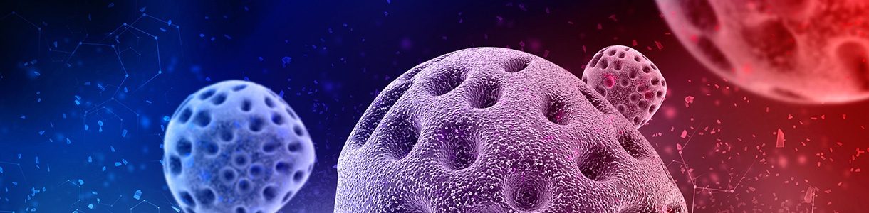
The Marburg and Ebola viruses are similar in their morphology, but differ in their antigenic structure. Polymorphism is characteristic, virions can be worm-shaped, spiral-shaped and rounded. Their length ranges from 665 to 1200 nm, the cross-sectional diameter is 70-80 nm. Ultrastructure and antigenic composition are different from all known viruses. Viral particles contain RNA, lipoprotein; the presence of hemagglutinins and hemolysins was not detected. Antigenic activity is associated with viral particles, the existence of a soluble antigen has not been proven. Viruses are isolated and passaged in guinea pigs and in a culture of transplanted green monkey kidney (Vero) cells. When passaged in tissue cultures, the virus has an incomplete cytopathic effect or does not cause it at all. It belongs to the family Filoviridae, the genus Lyssavirus.
The first outbreaks of the disease occurred in 1967 simultaneously. Marburg and Frankfurt am Main, one patient was observed at this time in Yugoslavia. The source of infection was mainly the tissue of African green monkeys (25 patients), there were also secondary diseases (6 patients) – from two doctors, one nurse, a morgue worker and the wife of a veterinarian. Of the 25 initially infected patients, 7 people died. Subsequently, similar diseases were observed in Sudan (area of the village of Maridi, the disease was called Maridi fever), in Kenya, South Africa. The source of infection and the reservoir of the virus in nature during all these outbreaks were African green monkeys (Ceropithecus aethiops), in which the infection can occur inapparently. The participation of other animals in natural foci of infection, as well as the mode of transmission of infection to monkeys has not yet been studied.
A sick person is a danger to others. Virus isolation occurs with the nasopharyngeal contents, urine, and the blood of patients is contagious. Infection in humans can occur through airborne droplets, when a virus hits the conjunctiva, or on the skin (accidental needle pricks or cuts), the possibility of sexual transmission of infection is not excluded (the virus was detected in the seminal fluid). The virus in the body of the ill person can persist for up to 3 months.
Pathogenesis during Fever Marburg
Gates of infection are damaged skin, mucous membranes (oral cavity, eyes). Characterized by dissemination of the virus. Reproduction can occur in various organs and tissues (liver, spleen, lungs, bone marrow, testicles, etc.). The virus is long detected in the blood, semen (up to 12 weeks). Histopathological changes are noted in the liver (liver cell obesity, necrobiosis of individual cells, cell infiltration), kidney (damage to the epithelium of the renal tubules), spleen, myocardium, lungs. Multiple minor hemorrhages in various organs (brain, etc.).
Fever Marburg Symptoms
The incubation period is 2-16 days. The clinical symptoms, severity and outcomes of the diseases described as Marburg fever and Maridi hemorrhagic fever are no different. Prodromal period is absent. The disease begins acutely with a rapid rise in body temperature to a high level, often with chills. From the first days of the disease, there are signs of general intoxication (headache, weakness, muscle and joint pain), after a few days lesions of the gastrointestinal tract, hemorrhagic syndrome join; dehydration develops, consciousness is disturbed.
In the initial period, the patient complains of a headache of a diffuse nature or more pronounced in the frontal region, stitching chest pains, aggravated by breathing, chest pains, sometimes a dry cough. There is a feeling of dryness and sore throat. There is hyperemia of the pharyngeal mucosa, the tip and edges of the tongue are red; vesicles appear on the hard and soft palate, tongue, at the opening of which surface erosions are formed; in contrast to Lassa fever, marked necrosis is not observed. The tone of the muscles, especially the back, neck, chewing muscles is increased, their palpation is painful. From the 3-4th day of illness, pain in the abdomen of a cramping character joins. The stool is liquid, watery, half of the patients have blood in the stool (sometimes clots) or signs of gastrointestinal bleeding (melena) are observed. Some patients develop vomiting with an admixture of bile and blood in the vomit. Diarrhea occurs in almost all patients (83%), lasts about a week; vomiting is less common (68%), lasts 4-5 days.
In half of the patients on the 4th-5th day of the disease, a rash appears on the body (sometimes core-like), in some patients, vesicular elements may occur on the background of a maculopapular rash. The rash spreads to the upper limbs, neck, face. Sometimes itchy skin. With the development of hemorrhagic syndrome, hemorrhages appear in the skin (in 62% of patients), in the conjunctiva, the oral mucosa. At this time, nasal, uterine, gastrointestinal bleeding appears. At the end of the 1st, sometimes on the 2nd week, the signs of toxicosis reach maximum severity. Symptoms of dehydration, infectious and toxic shock. Sometimes there are convulsions, loss of consciousness. During this period, patients often die.
In the study of blood marked leukopenia, thrombocytopenia, anisocytosis, poikilocytosis, basophilic granularity of erythrocytes. Cerebrospinal fluid remains unchanged even in patients with signs of irritation of the meninges. The recovery period is delayed for 3-4 weeks. At this time, there is alopecia, intermittent abdominal pain, loss of appetite and prolonged mental disorders. Late complications include transverse myelitis and uveitis.
Complications
Perhaps the development of early encephalitis, as well as myelitis, orchitis, mental disorders, reduced intelligence. In severe cases, infectious-toxic shock, hypovolemic shock, edema of the lungs and brain can serve as the causes of a lethal outcome.
The mortality rate reaches 30% or more, death usually occurs between the 8-17th day of the disease from hemorrhagic manifestations.
Diagnostics of Marburg Fever
In recognizing the disease, epidemiological prerequisites are important (being in areas with natural foci of Marburg fever, working with tissues of African monkeys, contact with patients). The clinical picture is characteristic: acute onset of the disease, severe course, presence of vesicular-erosive changes of the oral mucosa, hemorrhagic syndrome, exanthema, diarrhea, vomiting, dehydration, severe damage to the central nervous system (consciousness disorders, meningeal syndrome), characteristic changes in peripheral blood. The lack of effect from the use of antibiotics, chemotherapeutic and antimalarial drugs, the negative results of conventional bacteriological and parasitological studies have some significance.
Specific methods of laboratory studies can identify the virus or antibodies to it. Work with vaccinated material is carried out in compliance with preventive measures only in specially equipped laboratories. When taking material for laboratory research, observe the rules of packing and shipping recommended for especially dangerous infections (placed in metal tubes, sent to laboratories by hand). Antibodies in the serum of patients are determined using the immunofluorescence method.
Fever Marburg Treatment
Means etiotropic therapy are absent. Carry out pathogenetic and symptomatic therapy, injected plasma convalescents. The use of interferon and their inductors is ineffective.
Preventing Marburg Fever
Identification of patients, their isolation, quarantine measures. Heterogeneous (equine) serum immunoglobulin for immunoprophylaxis of high-risk contingents has been developed.