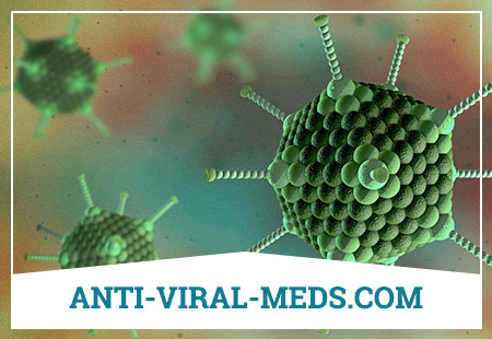
Adenovirus infection is an acute anthroponotic viral infection that affects the mucous membranes of the upper respiratory tract, eyes, intestines, lymphoid tissue and proceeds with moderate intoxication.
Brief historical information
Human adenoviruses were first isolated by W. Rowe (1953) from the tonsils and adenoids of children, and then from patients with acute respiratory viral infections and atypical pneumonia with conjunctivitis (Huebner R., Hilleman M., Trentin J. et al., 1954). In experiments on animals, the oncogenic activity of adenoviruses has been proven (Trentin J. et al., Huebner R. et al., 1962).
Etiology
The causative agents are DNA-genomic viruses of the Mastadenovirus genus of the Adenoviridae family. Currently, about 100 virus serovars are known, more than 40 of them have been isolated from humans. Serovars of adenoviruses differ sharply in epidemiological characteristics. Serovars 1, 2 and 5 cause lesions of the respiratory tract and intestines in young children with prolonged persistence in the tonsils and adenoids, serovars 4, 7, 14 and 21 – SARS in adults. Serovar 3 causes the development of acute pharyngoconjunctival fever in older children and adults, several serovars cause epidemic keratoconjunctivitis. Disease outbreaks are more commonly associated with types 3, 4, 7, 14 and 21.
According to the ability to agglutinate erythrocytes, adenoviruses are divided into 4 subgroups (I-IV). Adenoviruses are stable in the external environment, persist for up to 2 weeks at room temperature, but die from exposure to ultraviolet rays and chlorine-containing drugs. They tolerate freezing well. In water at 4 ° C, they remain viable for 2 years.
Epidemiology
The reservoir and source of infection is a person, a patient or a carrier. The causative agent is excreted from the body with the secret of the upper respiratory tract until the 25th day of illness and more than 1.5 months – with feces.
The mechanism of infection transmission is aerosol (with droplets of saliva and mucus), the fecal-oral (alimentary) route of infection is also possible. In some cases, the transmission of the pathogen is carried out through contaminated objects of the external environment.
The natural susceptibility of people is high. The transferred disease leaves type-specific immunity, repeated diseases are possible.
Main epidemiological signs. Adenovirus infection is ubiquitous, accounting for 5-10% of all viral diseases. The incidence is recorded throughout the year with an increase in cold weather. Adenovirus diseases are observed both in the form of sporadic cases and in the form of epidemic outbreaks. Epidemic types of viruses (especially 14 and 21) cause large outbreaks of diseases among adults and children. Adenovirus hemorrhagic conjunctivitis often occurs when infected with a virus of types 3, 4 and 7. The development of cases of conjunctivitis is associated with a previous respiratory adenovirus infection or is the result of infection with the virus through water in swimming pools or open water. Children of early age and military personnel are more often ill. The incidence is especially high in newly formed groups of children and adults (in the first 2-3 months); the disease proceeds as SARS. In some cases, nosocomial infection is possible during various medical procedures. The disease in newborns and young children proceeds according to the type of keratoconjunctivitis or damage to the lower respiratory tract. Rare adenoviral lesions include meningoencephalitis and hemorrhagic cystitis, more often detected in older children.
SARS, including influenza, constitute a complex of conjugate infections, so the process of spreading these infections is a single balanced system. Currently, about 170 types of pathogens are known to cause influenza-like diseases, and even during an epidemic, influenza accounts for no more than 25-27% of all acute respiratory viral infections.
Pathogenesis
In case of aerosol infection, the pathogen enters the human body through the mucous membranes of the upper respiratory tract and spreads through the bronchi to their lower sections. The entrance gates of infection can be the mucous membranes of the eyes, as well as the intestines, where the virus enters when swallowing mucus from the upper respiratory tract. The virus is localized in the cells of the epithelium of the respiratory tract and small intestine, where it multiplies. In the lesions, an inflammatory reaction develops, accompanied by an expansion of the capillaries of the mucous membrane, hyperplasia of the submucosal tissue with infiltration by mononuclear leukocytes and sometimes hemorrhages in it, which is clinically manifested by tonsillitis, pharyngitis, conjunctivitis (often membranous), and diarrhea. Sometimes keratoconjunctivitis develops with clouding of the cornea and visual impairment. By the lymphogenous route, the pathogen penetrates into the regional lymph nodes, where hyperplasia of the lymphoid tissue and the accumulation of the virus occur during the incubation period of the disease. In the clinical picture, these mechanisms cause the development of peripheral lymphadenopathy and mesadenitis.
As a result of suppression of macrophage activity and increased tissue permeability, viremia subsequently develops with dissemination of the pathogen in various organs and systems. During this period, the virus penetrates the vascular endothelial cells, damaging them. In this case, a syndrome of intoxication is often observed. Fixation of the virus by macrophages in the liver and spleen is accompanied by the development of changes in these organs with an increase in their size (hepatolienal syndrome). Viremia and reproduction of the pathogen in the cells of the epithelium and lymphoid tissue can be prolonged.
Clinical picture
The duration of the incubation period varies from 1 day to 2 weeks, more often 5-8 days. The disease begins acutely with the development of mild or moderate symptoms of intoxication: chills or chilling, mild and intermittent headache, myalgia and arthralgia, lethargy, adynamia, loss of appetite. From the 2-3rd day of illness, body temperature begins to rise, more often it remains subfebrile for 5-7 days, only sometimes reaching 38-39 ° C. In rare cases, pain in the epigastric region and diarrhea are possible.
At the same time, symptoms of damage to the upper respiratory tract develop. Unlike the flu, moderate nasal congestion appears early with abundant serous, and later – serous-purulent discharge. Sore throat and cough are possible. After 2-3 days from the onset of the disease, patients begin to complain of pain in the eyes and profuse lacrimation.
When examining patients, facial hyperemia, scleral injection, and sometimes a papular rash on the skin can be noted. Often develops conjunctivitis with hyperemia of the conjunctiva and mucous, but not purulent discharge. In children of the first years of life and occasionally in adult patients, membranous formations may appear on the conjunctiva, swelling of the eyelids increases. Possible damage to the cornea with the formation of infiltrates; when combined with catarrhal, purulent or membranous conjunctivitis, the process is usually unilateral at first. Infiltrates on the cornea resolve slowly, within 1-2 months.
Conjunctivitis can be combined with manifestations of pharyngitis (pharyngoconjunctival fever).
The mucous membrane of the soft palate and the posterior pharyngeal wall is slightly inflamed, may be granular and edematous. The follicles of the posterior pharyngeal wall are hypertrophied. The tonsils are enlarged, loosened, sometimes covered with easily removable loose whitish coatings of various shapes and sizes. Note the increase and pain on palpation of the submandibular, often cervical and even axillary lymph nodes.
If the inflammatory process of the respiratory tract takes on a descending character, the development of laryngitis and bronchitis is possible. Laryngitis in patients with adenovirus infection is rarely observed. It is manifested by a sharp “barking” cough, increased pain in the throat, hoarseness of voice. In cases of bronchitis, the cough becomes more persistent, hard breathing and scattered dry rales in different departments are heard in the lungs.
The period of catarrhal phenomena can sometimes be complicated by the development of adenovirus pneumonia. It occurs after 3-5 days from the onset of the disease, in children under 2-3 years old it can begin suddenly. At the same time, the body temperature rises, the fever takes on an irregular character and lasts for a long time (2-3 weeks). The cough becomes stronger, general weakness progresses, shortness of breath occurs. Lips become cyanotic. When walking, shortness of breath increases, perspiration appears on the forehead, cyanosis of the lips intensifies. According to radiological signs, pneumonia can be small-focal or confluent.
In young children, in severe cases of viral pneumonia, a maculopapular rash, encephalitis, and foci of necrosis in the lungs, skin, and brain are possible.
Pathological changes in the cardiovascular system develop only in rare severe forms of the disease. Muffled heart sounds and a soft systolic murmur at its apex are characteristic.
Lesions of various parts of the respiratory tract can be combined with disorders of the gastrointestinal tract. Abdominal pain and bowel dysfunction occur (diarrhea is especially common in young children). The liver and spleen are enlarged.
